RSID Kits
Introducing the newest colors for RSID cassettes. New look – same specificity and sensitivity – easily identifiable
The RSID Tests for Semen, Saliva, Blood and Urine are now color coded. This enables more rapid allocation of the test chips on the laboratory bench. Mix-ups and mistakes are thus virtually excluded.

Reduced Incubation Time
with no loss of sensitivity or specificity
To determine if RSID-Semen and RSID-Saliva are compatible with shorter sample extraction times, a series of time course experiments were undertaken with control swabs, aged samples (several years old). These data clearly demonstrated that similar results could be obtained from all tested sample types using incubation times as short as 10 seconds (with shaking/Vortexer) to as long as 1 hour.
Room temperature extraction of forensic samples for a minimum of 10 seconds with shaking is sufficient for detection of semenogelin with RSID-Semen and RSID-Saliva. Longer incubation times are optional.

RSID – Multiple Tests from one Piece of evidence
The Rapid Stain Identification (RSID) tests are designed for fast, easy, and reliable detection of human saliva, semen, blood and urine from a
variety of samples: bottles, cans, envelopes, plastic straws, bite marks, vaginal swabs and stained surfaces etc. No inhibition
by the presence of other body fluids – blood, semen, vaginal secretions, or urine. The tests are Lateral Flow Strip Tests.
RSID-Semen and RSID-Blood are CONFIRMATORY for the detection oh human semen respectively blood.
PRINCIPLE OF THE IMMUNOCHROMATOGRAPHIC FLOW TESTS:
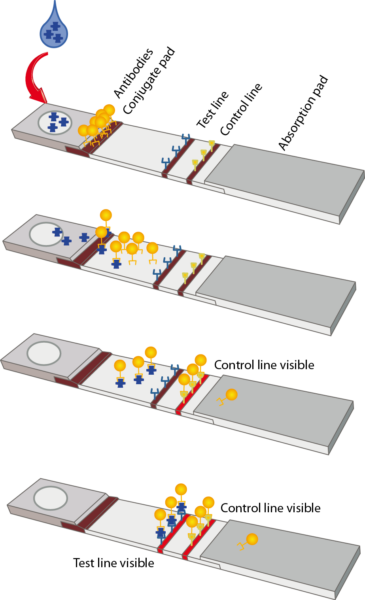
The RSID Saliva test for example is an immunochromatographic assay that uses two monoclonal antibodies specific for human salivary α-amylase. This system consists of overlapping components treated such that the tested fluid is transported from the conjugate pad to the membrane and
is finally retained on the wick. The conjugate pad and membrane are pretreated before assembly. The user just needs to add the extract in diluent buffer (provided) to initiate the test. Once the tested sample is added to the sample window, the buffer and sample diffuse through the conjugate pad, which has pre-dispersed colloidal gold conjugated anti-human salivary α-amylase monoclonal antibodies. The diluent redissolves the colloidal gold labeled anti α-amylase antibodies
which will bind salivary amylase if it is present in the sample. Salivary α-amylase-colloidal gold antibody complexes are transported by bulk fluid flow to the membrane phase of the test strip.
These complexes, if present, migrate along the membrane and are bound at the ‘test line’ creating a visible red ‘bar’ against the white membrane. Colloidal gold labeled mouse antibody will continue to progress along the membrane and be bound by anti-mouse antibody at the ‘control line’ again creating a visible signal. A visible line at the ‘test’ position indicates the presence of human saliva; a visible line at the ‘control’ position indicates that the test is working correctly.
